Bronchoscopy Hospital in Nashik – Dr. Mutha Chest & Sleep Center
Dr. Mutha Hospital is recognized as one of the Best Bronchoscopy Hospitals in Nashik, offering advanced bronchoscopy procedures for both diagnosis and treatment of various lung diseases. As a leading Bronchoscopy Hospital in Maharashtra, India, we utilize cutting-edge technology and maintain a patient-focused approach for accurate and safe results.
Instrument Used
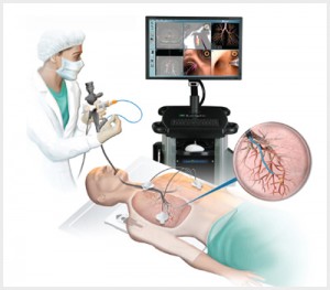
At Dr. Mutha Hospital, we use the latest Bronchovideoscope by OLYMPUS – BF 1TQ 170, ensuring top-notch imaging and procedure precision.
Key Features:
Excellent Image Quality: High-resolution imaging ensures accurate diagnosis.
NBI (Narrow Band Imaging): An advanced optical enhancement technology that improves the visualization of vessels on the mucosal surface.
Wider EndoTherapy Device Capability: The 2.8 mm instrument channel allows the use of various EndoTherapy devices, enhancing therapeutic possibilities.
How the Test is Performed
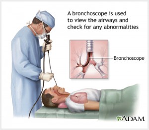
Bronchoscopy involves using a bronchoscope—a flexible tube—to view the inside of your lungs. This scope is typically inserted through the mouth or nose, passing down the windpipe (trachea) into the lungs. In most cases, a flexible scope is used. The procedure is usually done while you’re awake but sedated.
Medications are given intravenously to help you relax. A numbing anesthetic spray is applied to your mouth and throat. If the scope is inserted via the nose, a numbing jelly is used. Coughing may occur at first but reduces as the anesthesia takes effect.
The doctor may:
Perform a therapeutic lavage using saline to collect lung fluid or cells.
Use tiny brushes, forceps, or needles to perform a biopsy.
Use ultrasound to view lymph nodes or structures around the airways.
Place airway stents if needed.
How to Prepare for the Test
Before undergoing bronchoscopy at our Nashik facility, please:
Avoid food and drink for 6–12 hours before the procedure.
Stop blood thinners like aspirin or ibuprofen (consult your doctor).
Arrange for transportation to and from the hospital.
Plan for rest on the day of the procedure.
Most tests are performed on an outpatient basis, though some may require overnight observation.
How the Test Will Feel
A local anesthetic is used to numb your throat. As the numbing begins, fluid in the throat may cause coughing or gagging, which subsides soon. During the procedure, you may feel mild pressure or tugging. The relaxant medication ensures comfort and often results in no memory of the procedure.
Post-procedure, you may experience a scratchy throat. Cough reflex returns within 1–2 hours, after which you can eat or drink.
Why the Test is Performed
Our Bronchoscopy services in Nashik help diagnose or treat a wide range of lung conditions.
Common Diagnostic Reasons:
Suspicion of lung cancer, atelectasis, or lymph node involvement
Unexplained cough lasting over 3 months
Hemoptysis (coughing up blood)
Suspected interstitial lung disease
Foreign object in airway
Undiagnosed lung infections
Exposure to toxic gases
For diagnosis of Interstitial Lung Disease in Nashik, see our ILD page.
Therapeutic Uses:
Remove fluid or mucus plugs
Remove foreign objects
Widen blocked or narrowed airways
Drain abscesses
Administer lung cancer treatments
Perform therapeutic lavage
Normal vs Abnormal Results
Normal Results:
No foreign objects or blockages
Healthy lung cells and fluids
Abnormal Results May Indicate:
Bacterial, viral, fungal, or TB infections
Lung cancer
Sarcoidosis or vasculitis
Inflammatory lung diseases
Trachea or bronchi narrowing
For more on Pulmonary Tuberculosis treatment in Nashik, visit our TB Treatment page.
Risks
Though bronchoscopy is safe, potential risks include:
Minor bleeding
Infection
Rare complications: arrhythmias, low oxygen, fever, pneumothorax, sore throat, or heart attack (in high-risk cardiac patients)
We follow strict protocols to minimize all risks at our bronchoscopy unit in Nashik.
Why Choose Dr. Mutha Hospital for Bronchoscopy in Nashik?
NABH-accredited multispecialty facility
Advanced Olympus bronchoscopy system
Experienced Pulmonologist in Nashik
Clean, patient-first care
Integrated diagnosis with Pulmonary Function Tests (PFT) and Sleep Study Diagnosis.
Book Your Bronchoscopy Test in Nashik Today
For appointments with the Best Bronchoscopy Specialist in Nashik, contact Dr. Mutha Chest & Sleep Center today. Early diagnosis can prevent complications and help in managing critical respiratory illnesses.